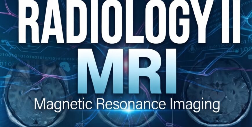
Radiology II. MRI For Physiotherapists

- Description
- Curriculum
- Reviews
Course Overview
MRI shows what X-ray and CT can’t — soft tissue, ligament, and marrow detail with zero radiation. This course takes you from the physics of how an MRI image is formed to reading real brain, spine, and knee scans with confidence.
Built to give you a repeatable method for reading any MRI — not a list of pathologies to memorize.
What You’ll Walk Away With
The ability to open an MRI report or scan and actually understand what it’s telling you — and why the radiologist chose that sequence in the first place.
Understand the “Why”
Why T1 looks different from T2, and why a radiologist picks one sequence over another for a given question.
Read With a System
A structured, repeatable method for scanning any MRI — brain, spine, or joint — so nothing gets missed.
Connect Findings to Care
Translate an imaging finding into what it actually means for your treatment plan.
From the Brain to the Knee
Brain MRI
Normal anatomy and the neurological findings you need to recognize — and the safety screens that come before every scan.
Lumbar Spine MRI
Reading disc and nerve root findings with a clear, step-by-step inspection method.
Knee MRI
Ligament, meniscus, and cartilage injuries — read with the same precision as the report on your desk.
Key Clinical Questions
A few examples of what you’ll be able to answer with confidence by the end:
- Why does this report start with the T2-weighted images?
- What does this finding actually mean for how I treat this patient?
- Which patients should never go near an MRI scanner?
Prerequisites
- Physical therapist or medical professional
- Recommended: complete Radiology I. X-ray first
What’s Included
- Certificate of completion
- Lifetime access